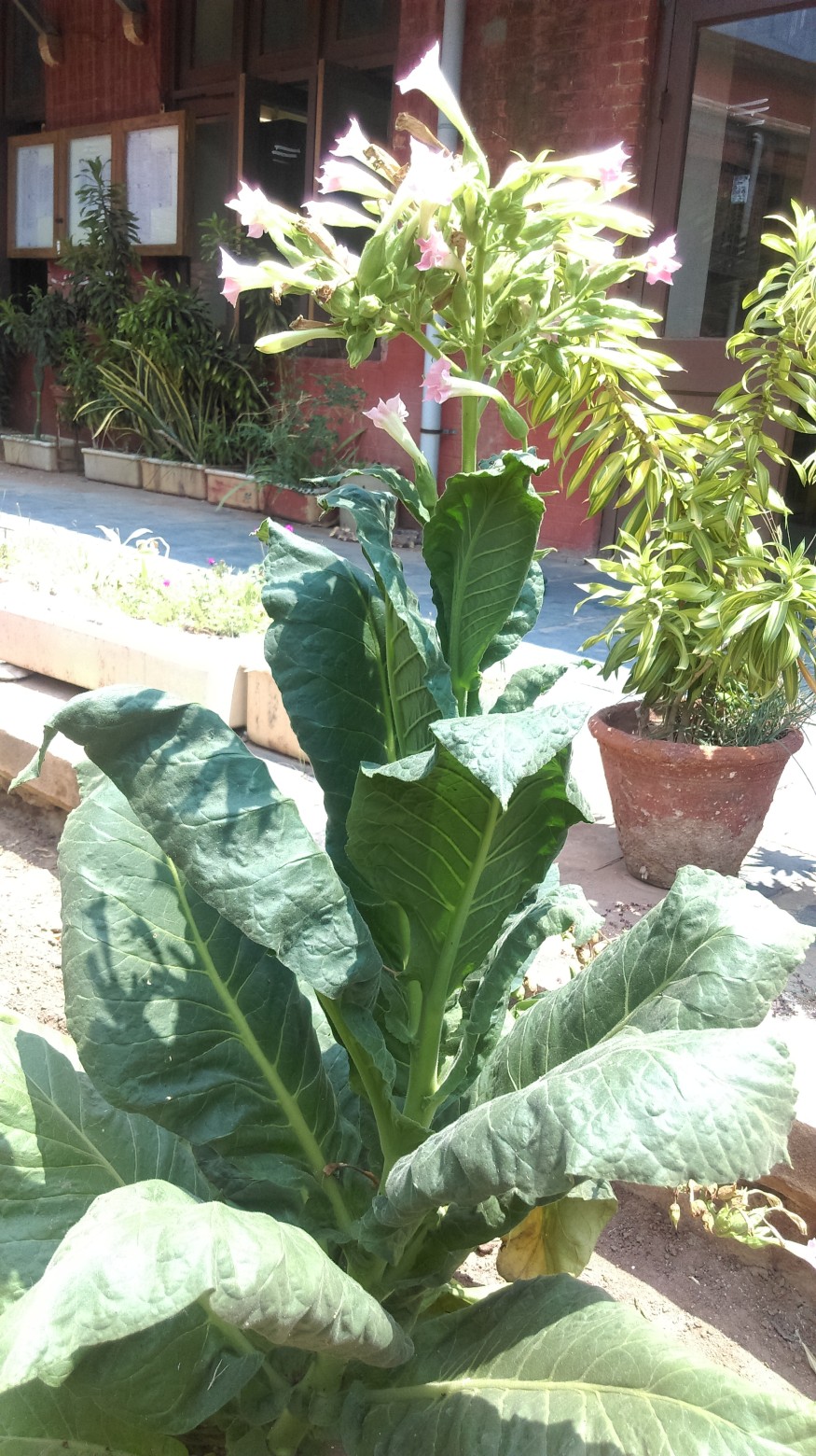
For survival, plants rely on their capacity to detect light. Plants, unlike mammals, do not have photoreceptors in their eyes to capture and transmit information from visual inputs.
Instead, plants are covered with a network of light-sensing photoreceptors that sense various wavelengths of light, allowing them to control their lifecycles and adapt to changing environmental conditions.

Research
Scientists from the Van Andel Institute and Washington University have finally discovered the molecular structure of one of these critical photoreceptors, PhyB, showing a completely different system. The discoveries published in the journal Nature might have ramifications for agricultural and "green" biotech methods.
According to VAI Professor Huilin Li, Ph.D., co-corresponding author of the study, photoreceptors like PhyB help plants sense and respond to the world around them by influencing life-sustaining processes like shade avoidance, seed germination, flowering time determination, and the development of chloroplasts, which convert light into usable energy.
"Our novel structure provides insight on how PhyB functions and has the potential for a wide range of applications in agriculture, renewable energy, and even cellular imaging," says the researcher.
Understanding the System

Understanding PhyB's shape is crucial because it directly influences how it interacts with other molecules to transmit light changes and helps plants adapt by causing changes in gene expression. Previous studies on PhyB only offered a truncated image of the complete molecule rather than a comprehensive depiction.
Li and research co-corresponding author Richard D. Vierstra, Ph.D., of Washington University, used one of the most studied plants on the planet, Arabidopsis thaliana, to create a near-atomic resolution picture PhyB. Because it reproduces fast, is tiny, and easy to manage, this little blooming plant is a good model for study.
Implications
The study team captured approximately 1 million particle pictures of PhyB coupled to its natural chromophore - a molecule that absorbs a specific hue of light - using VAI's high-powered cryo-electron microscope, or cryo-EM.
They then reduced the photos to 155,000, which they utilized to comprehensively represent PhyB's structure at a 3.3 Ångstrom near-atomic level.
Instead of the parallel structure reported in previous investigations, they discovered a sophisticated 3D structure with parallel and anti-parallel portions. The findings imply that PhyB may magnify modest changes in light-sensing chromophore molecules and dramatically modify its shape, signaling that light is available to the plant.
The finding is the culmination of a decade of work between Li and Vierstra. It completely changes our understanding of PhyB and phytochromes, the receptor family to which PhyB belongs. PhyB and other phytochromes were formerly thought to be identical to those found in single-celled organisms such as bacteria.
The new findings challenge that idea and establish the groundwork for more research into the nitty-gritty of PhyB and phytochrome activity.
Related Article : Flourishing Plants in Antarctica Signals Worsening Climate Crisis
For similar news, don't forget to follow Nature World News!
© 2025 NatureWorldNews.com All rights reserved. Do not reproduce without permission.


