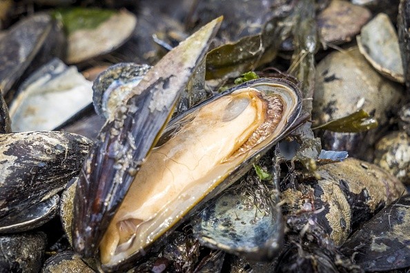
Those who have attempted to extricate a mussel from anything--from wood to rock--are well aware of how stubborn the aquatic bivalves are, and their gluey secret has long captivated researchers.
For years, scientists have sought to replicate the incredible adhesive and its properties in the lab, concentrating on a few of the eight proteins generated by mussels and used to bind an organ simply known as the foot, which mussels use to adhere to surfaces.
The inspiration of a synthetic cement

According to ScienceDaily, Northwestern University researchers generated a substance that outperforms the glue they were attempting to emulate by employing a unique approach of arranging molecules.
Their findings, which is published on March 3 in the Journal of the American Chemical Society, go into further detail about how these protein-like polymers might be used as a platform to build innovative materials and therapeutics.
According to Northwestern's Nathan Gianneschi Professor of Chemistry at the Weinberg College of Arts and Sciences, the polymer might be employed as an adhesive in a biological environment, which implies it could now be stuck to a specific tissue in the body.
Or Berger, a postdoctoral researcher at Gianneschi's lab who researches peptides, is the paper's first author.
Instead of exactly replicating the structure of mussel proteins, these precise chains of amino acids developed an idea for how to assemble amino acid basic components differently in order to mimic the properties.
Berger reasoned that adding a fundamental foundation of one of the enzymes into the synthetic polymer would improve its properties.
The 'gluey' secret of mussels
Mussels spend much of their time surviving the harsh intertidal habitat and smashing seas.
How they manage to stay tied to rocks and their fellow mussels in these settings has long captivated experts and students alike, as per The McGill Tribune.
Evolution, fortunately, provides solutions to such challenging and complex design challenges, as well as inspiration for human engineers.
A revolutionary McGill-led study published in Science shows how mussels create their very strong adhesive.
The scientists used micro-CT tomography to rebuild the mussel foot of the blue mussel Mytilus edulis, showing a complex system of longitudinal ducts (LDs), or long tubes.
These LD microchannels, according to two distinct imaging methods, are formed by a swarm of small hairs known as cilia and microvilli.
Through an acidic discharge of liquid protein precursor into the distal depression, an indentation at the tip of the foot's ventral side, the mussels release byssus fibers, which adhere the mussel to a solid surface.
The iron and vanadium ions required to create the plaque collect and are stored in the foot, away from the proteins required for the glue.
During plaque formation, tiny metal specks known as cytoplasmic metal storage particles (MSPs) are transported and combined.
The MSPs release metal ions that distribute throughout the plaque as the secreted contents pass through the channels, allowing the glue to preserve its structure.
© 2025 NatureWorldNews.com All rights reserved. Do not reproduce without permission.


