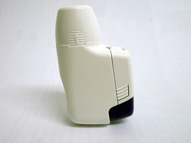
Researchers have made new inroads into understanding aerosol treatments for asthma to help in improvements of the drug in the future. Such innovation can potentially help asthma patients.
Asthma is a disease of the lungs that affects around 330 million people all over the world. It is a major worldwide health concern. Presently, the best treatment form and preparation available with the most beneficial impact are aerosols inhaled directly into the patient's lungs. It is a challenge for researchers because our current knowledge regarding the microstructure of drugs before being converted to aerosol is quite limited.
Such aerosol preparations are carrier-based DPI or dry powder inhalers, which must be characterized accurately for particle size distribution, surface roughness, flow properties, and fines contents. It is critical to understand the powder formulation's micro-structure, but before the current research, characterization techniques have not given complete information.
Conventional techniques include optical microscopy or OM and laser diffraction or LD. These techniques are limited because the particles are assumed to be spherical and because they provide varying results based on particle dispersion and orientation.
Scientists from the University of Manchester have done research that sheds light on this aspect. Using X-Ray Micro-Computer Tomography (or XCT) scanning, the microstructure of the drug's particles have been quantified to the nano-level or scale.
Researchers revealed this 3D microstructure for the first time. It gives the scientific community, medical researchers, and pharmaceutical companies more understanding regarding the drug's behavior when it is aerosolized.
Dr. Parmesh Gajjar, the research's lead author, said that the team was successful in visualizing a 3-D drug-blend to see the interaction between its active ingredient and its non-drug particles. Such insight is essential in the final step of quality control of drug production. It will enable producers to verify the actual drug content in the medicine and help in coming up with better formulations, and thus improve its effectiveness.
The XCT equipment and instruments that the research team utilized is located at the world-leading HMXIF or the Henry Moseley X-ray Imaging Facility. This facility is housed at the University of Manchester. The facility and equipment are capable of analyzing samples at a resolution as small as 50nm.
The research is very important in obtaining knowledge about asthma inhalation medicines, which need aerosolization for generating particles small enough to be absorbable by the lungs. The project studied particles that are tiny enough to be able to reach into the lung's deepest recesses.
The research's novel technological innovation was one of the papers proposed to be presented at the 2020 Respiratory Drug Delivery conference (RDD); it was chosen one of the key presentations at the said global conference. The venue of the conference was initially booked at Palm Springs. Still, due to the coronavirus pandemic and the lockdowns it caused, the organizers first opted to make it an online event instead and has currently been canceled until further notice.
The study was published in the European Journal of Pharmaceutics & Biopharmaceutics and is entitled "3D Characterisation of Dry Powder Inhaler Formulations: Developing X-Ray Micro Computed Tomography Approaches.
© 2025 NatureWorldNews.com All rights reserved. Do not reproduce without permission.


